

How To Make A Neuron Model

 Beady Neuron
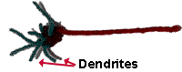
Become out those chaplet and make a neuron! This neuron with seven dendrites requires 65 beads: 42 beads for the dendrites, x beads for the cell body, 12 beads for the axon and 1 dewdrop for the synaptic terminal. String the chaplet using the blueprint in the diagrams below. The string can be yarn, rope, or for the best result use flexible wire. Yous can also create your own pattern or use a dissimilar colored bead for a nucleus in the jail cell torso.
 Materials:
- Wire
- 65 beads
- or get a full Beady Neuron Kit

Pipe Cleaner Neuron
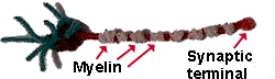
Go out those pipe cleaners and brand a neuron! This neuron pipe cleaners of 5 different colors: one colour each for the dendrites, jail cell torso, axon, myelin sheath and synaptic final. Any colors will do.
| | 1. Have one pipe cleaner and roll it into a ball. This is will be the jail cell trunk. 2.Take another piping cleaner and adhere it to the new "cell body" by pushing it through the ball so there are two halves sticking out. Take the two halves and twist them together into a single extension. This will be the axon. |
 | 3.Accept other pipe cleaners and push them through the "cell trunk" on the side reverse the axon. These are dendrites. These can be shorter than your axon and you tin can twist more pipage cleaners to make more dendrites. |
 | iv.Wrap modest individual pipage cleaners along the length of the axon. These volition represent the myelin sheath. five. Wrap another pipe cleaner on the finish of the axon. This volition exist the synaptic concluding. |
Cord Neuron
 Information technology'due south a parachute! Information technology's a witch'south broom! It'south the Eiffel Tower! No, information technology's a NEURON!!!
If yous have ever played any "string games," then this neuron model should be like shooting fish in a barrel for y'all to brand. Follow the steps on this page to make a neuron from string.
Materials:- A loop of string or yarn (about 3 ft. in length).
Rope Neuron
Set upwardly the model:
- Get volunteers to agree each of the dendrites.
- Become ane volunteer to hold the cell body and one to concur the synaptic terminal. Make sure the person belongings the synaptic terminal keeps his or her hands AWAY from the place the axon attaches (more well-nigh this later).
- Get i volunteer who will hold more molecules of neurotransmitter (more plastic balls) most the people who are dendrites.
- Get one volunteer to hold the action potential.
Use the model:
- Have the person holding molecules of neurotransmitter TOSS the plastic balls to the people who are dendrites. The "dendrite people" try to catch the plastic assurance. This models the release of neurotransmitters and the attachment (binding) of neurotransmitters to receptors on dendrites.
- When three plastic balls are defenseless by dendrites, the person belongings the activeness potential can throw/slide the puddle float downward the axon. This simulates the depolarization of the neuron above its threshold value and the generation of an action potential.
- The action potential (puddle bladder) should speed down the axon toward the synaptic terminal where it will slam into the container. This should cause the release of the neurotransmitters (plastic balls) that were existence held there.
 CAUTION: The pool float will travel very fast! Make sure that the person belongings the synaptic terminal keeps his or her fingers and easily Away from the puddle float.
If the unabridged model is stretched tightly, the pool bladder should travel down to the terminal smoothly. This model tin can be used to reinforce the "ALL-OR-NONE" concept of the action potential:
- Once the activeness potential starts, it continues without interruption.
- The size of the action potential stays the same as it travels down the axon.

 Materials:
- Rope (for dendrites and axon)
- Plastic containers (for cell body and synaptic last)
- Puddle Float (or another object will slide along the rope; for the action potential)
- Plastic assurance (for neurotransmitters)
- Volunteers!
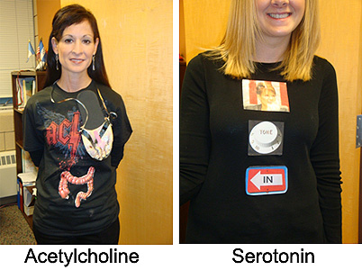
Neuron Costume
 Tin't think of a costume for Halloween? Why not be a neuron? The thought sent in past Kate Five.; y'all can encounter her is wearing her neuron costume in the photograph.)
Cut some piping cleaners into short pieces. Wrap these short pieces effectually longer pipe cleaners to make dendrites. Wrap one finish of each dendrite around a rubber pin. Pin the dendrites to a pink brusk-sleeved shirt and hat. Put on your costume...be a neuron!
Materials:
- Pinkish short-sleeved shirt
- Blueish long sleeved shirt
- Pink shorts
- Blue tights
- Blue hat
- Safety pins
- Blue pipe cleaners
Neuron...in a Pocketbook!
An edible neuron? Mix i box of Clot-O with water by following the directions on the Clot-O box. Afterward the Jell-O has cooled to a warm temperature, pour it into small plastic bags. Add fruits (canned fruit cocktail works well) and candies to the Jell-O to represent the organelles y'all would find within of a neuron. For example, standard mandarin orange slices could be mitochondria; a cherry half could be the nucleus; red and black cord licorice could be microtubules and neurofilaments. The plastic handbag can correspond the jail cell membrane. Don't forget ribosomes, the golgi apparatus and endoplasmic reticulum. You lot should also brand a "fable" of your cell then you remember which food represents which organelle. Write your legend on some card stock or index card. Afterward all the "organelles" have been added, tie off the top of the bag with a twist necktie and place the "cell" in the refrigerator. When the Jell-O gets firm, take it out, and compare your neuron to other neurons. So, have a snack...a neuron snack.
Materials:
- Jell-O - any season
- Plastic numberless - sandwich size
- Canned fruit
- Candies
- Twist ties
- A picture or diagram of a neuron
Run into cells of the nervous organization for more than virtually the organelles found in neurons.
Simple Neuron Model
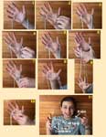
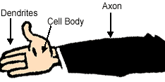
Here's the well-nigh simple model of a neuron I tin can think of...and you don't demand any supplies. It's your mitt! Hold out your arm and spread your fingers. Your paw represents the "cell body" (also called the "soma"); your fingers stand for "dendrites" bringing data to the prison cell body; your arm represents the "axon" taking data away from the prison cell body.
Materials: NONE
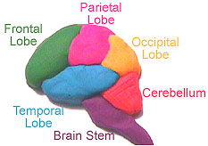
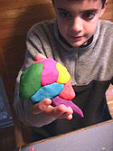
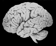
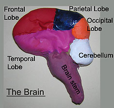
Model a Brain
Create a model of the encephalon by using clay, playdough, styrofoam, recyclables, food, etc. Create a whole brain or employ a encephalon atlas and create cross-sections of the brain at unlike levels. Use different colors to bespeak dissimilar structures.
Materials:
- Clay or Playdough or Styrofoam or Recyclables (canteen caps, cups, buttons, etc) OR Food (fruit, jelly beans)
- A film or diagram of the brain
Encephalon "Recipes"
Here are ii recipes for the structure of a model encephalon:
Recipe 1 (from the Pacific Science Center and the Group Wellness Cooperative in Seattle, WA)
Materials:- 1.five cups (360 ml) instant potato flakes
- 2.5 loving cup (600 ml) hot water
- 2 cups (480 ml) clean sand
- 1 gallon ziplock handbag
Recipe two (from BrainLink)
Materials:- two cups water
- 2 cups flour
- 4 teaspoons cream of tartar
- I quarter cup vegetable oil
- 1 cup salt
- Red food coloring
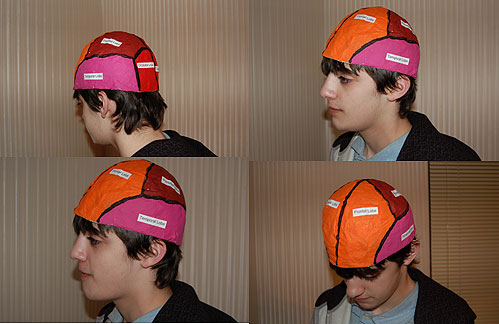
Thinking Cap
Display your brain on a "Thinking Cap." Thinking Caps are created from papier (or paper) mache.
Create the Form: First, create the brain course for the cap. Y'all tin create a class from wire (e.1000., craven wire) or a balloon or use a bowl to build your cap around. You could even ball up some newspaper and cover it will masking tape. The form should have the guess size and shape of your head so you lot tin can clothing it.
Create the Structure: Cut strips of newspaper and glue them to the form using papier mache paste. Pastes can exist made from:
- White gum and water (about 2 parts glue to one part h2o)
- White flour, salt and h2o (virtually i function flour to one part water with a few tablespoons of common salt)
- Liquid starch
Decorate the Thinking Cap: yous can paint the Thinking Cap with the lobes of the brain (see photo) or with the different areas of the cerebral cortex.
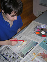
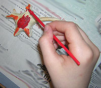
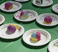
Baked Brains/Baked Neurons
Baked brains and neurons may look and smell tasty, but don't eat them. Mix flour and salt in a large bowl. Add water and mix. The mixture should start to stick together. If the mixture is too crumbly, add a little more water.
Spread a little flour on a countertop or cutting board. Work the mixture into a ball and knead it on the countertop or cutting board. When the mixture can be molded, have pieces and shape them into brains or neurons.
Place the finished brains and neurons on an ungreased cookie sheet. Bake in the oven at 350o for 10-fifteen minutes. The brains and neurons will turn slightly brown, but don't let them burn down. Let the brains and neurons cool, then paint them.
CAUTION: Be extremely careful using the oven. The cookie canvas and baked items tin get VERY hot. Adult supervision is required!
Materials:
- Flour (1 cup)
- Salt (1/iv loving cup)
- Water (1/3 to 1/2 cup)
- Oven for baking
- Paints
| Baked Brains/Broiled Neurons | ||
Uncooked Neuron | Broiled Neuron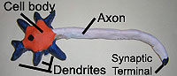 | Baked CNS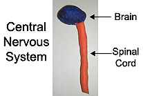 |
 |  | Uncooked CNS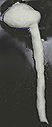 |
 | Uncooked Brain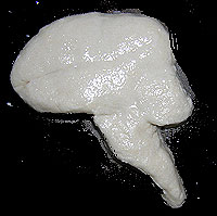 | Broiled Neurons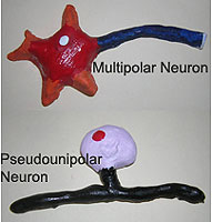 |
Practise You Know Your Brain?
 Grades 1-12
Alexandra Colón Rodriguez, a PhD student in Comparative Medicine and the Integrative Biology Program, Ecology and Toxicological Sciences Program at Michigan State University, has created a great hands-on activeness to learn about the brain.
Know Your Brain Activity
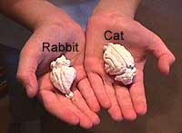
Make a Cat and Rabbit Encephalon
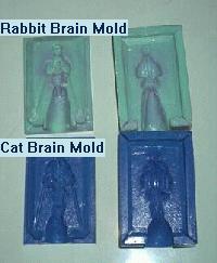 Grades four-12
Make brains again and again. BrainLink has developed cat and rabbit brain molds that yous can buy from the Carolina Biological Supply Visitor (CBS) for $16.95 each (Catalog #MF-95-2849A) . Coat each side of the rubber mold with liquid hand soap. Mix up FAST set dental plaster (also bachelor from CBS) with h2o to the consistency of toothpaste. Pour the dental plaster into each side of the mold. Sandwich the mold together and wait almost 15-20 minutes. Tap the mold a few times to get out all the air bubbles. Information technology can go a chip messy. When the plaster has set and is hard, skin dorsum i side of the mold and remove the brain. You can add together food coloring to the plaster while you are mixing the plaster if yous desire a brain with a bit of color or you can paint the different parts of the brain with different colors.
Materials:
- Brain Molds
- Fast set up dental plaster (telephone call a local dental supply company - it is fairly cheap - about $15 for 25 pounds - enough for many brains). Patterson Dental Supply, Inc. too has the plaster (catalog #48512). Their phone number is 1-800-626-5141 or ane-502-459-7444.
- Food coloring and paint (if you lot want to color the brains)
- Water - to mix up the plaster
Jello Brain
 Grades K-12
Get jello molds in the shape of the brain at Archie McPhee. For about $12 (plus shipping) you get either a gelatin mold of the top one-half of the brain or a side (lateral) view of the brain. Make brains over and once more. You tin can also model the meninges (coverings) of the brain past using layers of plastic wrap on peak of your jello brain. Make sure anybody gets a taste. Now that's what I call brain nutrient!
Hither is the recipe for the acme view jello brain:
- iii large (half dozen oz) boxes of jello (peach or watermelon recommended)
- 1 can (12 oz) evaporated skimmed/fat-gratis milk
- A few drops of green food coloring (to change the color to gray)
- iii.5 cups of water (two.5 cups boiled; ane cup common cold)
- Glaze mold with vegetable oil or spray
- Add 2.5 cups of boiling water into jello. Stir and deliquesce jello.
- Stir in 1 loving cup of common cold water.
- Stir in skimmed milk (~2 minutes)
- Add a few drops of greenish food coloring
- Pour entire mixture into jello mold
- Place mold into refrigerator overnight.
Make the Bones of the Spinal Column (Vertebrae)
 The homo spinal string is protected by the bony spinal column shown. There are 31 segments of the spinal cord and 33 bones (vertebrae) that environment these segments. There are 7 cervical vertebrae, 12 thoracic, 5 lumbar, 5 sacral and 4 coccygeal vertebrae in the human trunk. To model these bones, get 33 empty spools of thread (buttons may likewise work or slices of paper towel holders). Run a string or thread through the center of one of the spools or buttons. Tie off one finish of the string and put the remaining spools or buttons on the string. Each spool (or button) volition represent 1 vertebra. When your model is finished, discover how it can bend. In a real spinal cavalcade, the vertebrae are held together by ligaments.
Materials:
- Empty thread spools or buttons
- Cord
Read more most the spinal column.
Cap Caput...No, information technology'due south your Encephalon!
A great way to introduce the brain. Get a white swimming cap - you know, the kind that pulls on tight over your caput. Draw an outline of the brain on the cap with a black marker. To introduce the encephalon to your class, wear the cap!! It is a corking way to start a discussion. You could also depict the lobes of the brain or different areas of the cerebral cortex on your cap with different color markers.
Materials:
- White Swim Cap
- Black Marking
- Color Markers
Connect the Dots
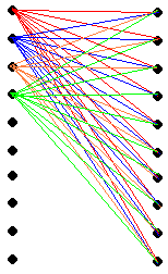 This exercise is to illustrate the complexity of the connections of the brain. Draw 10 dots on i side of a piece of paper and 10 dots on the other side of the paper. Assume these dots correspond neurons, and assume that each neuron makes connections with the x dots on the other side of the paper. Then connect each dot on 1 side with the 10 dots on the other side. As you can see from the diagram below, information technology gets very complicated afterward a while. I take only connected 4 of the "neurons".
Recollect that this is quite a simplification. Each neuron (dot) may really make thousands of connections with other neurons. If you tried this your newspaper would be really messy!!
Materials:
- Pencil, pens, markers
- Paper
Compare and Contrast
What improve model of the brain than a REAL BRAIN!! Endeavour to get "loaner" brains (human and animal) from your local academy (try medical schools, Departments of Biology, Zoology, Psychology). Some animal supply companies also sell brains (come across the Resources Page). Y'all may be able to find moo-cow or pig brains at the supermarket or local butcher.
Endeavor to become a "Brain Atlas" or await at some pictures of the brains here at Neuroscience for Kids or visit the Mammalian Brain Collection at the Academy of Wisconsin. This will aid the identification of brain structures.
Make certain you wear gloves when handling whatsoever specimens. Also be aware that some brains may exist perserved with formaldehyde solutions which have an unpleasant aroma and also should be handled with care.
Afterwards you lot accept collected all the specimens:
 Compare and Discuss:
- What are the similarities and differences between the brains?
- What are their relative sizes?
- Identify areas of the brain. Cortex? Cerebellum? Cranial nerves?
- Are their noticeable differences in whatever particular parts of the brains?
- Is the cortex smooth or rough?
- Compare placement of the cerebellum and spinal string.
- Compare size of olfactory bulb.
- Compare size of cerebral cortex.
- Discuss brain weight vs body weight issues.
- Hash out brain size and intelligence.
- Discuss language and brain size.
- Discuss cortical expansion in higher species.
- corpus callosum
- thalamus
- pons
- inferior and superior colliculus
- cingulate cortex
- medulla
- cerebellum
Materials:
- A brain
- A long pocketknife (this should only exist used within the lab)
- Trays (to agree encephalon specimens)
- Gloves (for handling specimens)
- Masks if the odor is strong
- Encephalon atlas
- Pointing devices (popsicle stick, probe, toothpick) to identify structures
Model a Retinal Image
 The encephalon has a tough job. It is works all the fourth dimension and the eye has to make things difficult. The convex nature of the lens of the centre turns an epitome upside downward on the retina. The brain must make sense of this and turn it "right-side upwardly". To model what a convex lens does to an prototype, get a magnifying drinking glass. Find a white wall or tape a white piece of paper to a wall that faces a window. Hold the magnifying glass close (3 in; ten cm) to the white wall or paper. You should see an inverted image of whatever is outside of the window. This is what is projected onto your retina.
Materials:
- Magnifying drinking glass
- White Wall or Paper and tape
Read more about the retina.
Message Transmission
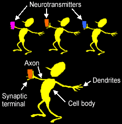 Messages can travel in neurons at speeds up to 268 miles/hr! These signals are transmitted from neuron (nerve cell) to neuron beyond "synapses."
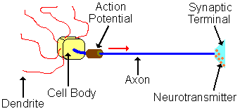
Let'southward make a concatenation of neurons...have everyone stand up and form a line. Each person in the line is a neuron. As shown in the figure on the right, your left hand are the dendrites of a neuron; your torso is the cell trunk; your right arm is an axon and your correct hand is the synaptic concluding. Your correct hand should have a small vial of liquid or some other detail, such as a push button or pebble, to represent neurotransmitters.
Each person should exist near arms length away from the next person. When the leader says "Get," take the person at the offset of the line start the signal manual by placing his or her "neurotransmitter" into the hand of the adjacent person. Once this message is received, this second neuron places its neurotransmitter into the dendrite of the next neuron. The third neuron then places its neurotransmitter into the dendrites of the next neuron and the "betoken" travels to the end of the line. The transmission is complete when the "signal" goes all the manner to the end of the line.
Call up that each "neuron" will pass its own transmitter to the adjacent neuron in line. Each neuron HAS ITS OWN neurotransmitter.
Allow'south review
- What are the parts of a neuron? The hand that receives the neurotransmitter is the "dendrite." The middle function of your body is the "soma" or "cell body." The arm that passes the neurotransmitter to the next person is the "axon" and the hand that gives the slap is the "synaptic terminal". In between the hands of 2 people is the "synaptic gap". For more than about the parts of a neuron, run into cells of the nervous system and the synapse.
- Mensurate how long it takes the message to go from the start neuron to the final. As well, measure the distance from the first to the last neuron. Now calculate the speed. How fast did the bulletin travel from kickoff to final neuron? Why do you lot think the speed of manual of the model is then slow?
Materials:- Stopwatch
- Vials for neurotransmitters
Saltatory Conduction
Saltatory conduction is a way that myelinated axons transmit action potentials. Activeness potentials bound from node to node. To model this, have everyone stand up upwardly and form a straight line. Each person should be at artillery length of the adjacent person. Requite the concluding person in line a pocket-sized object like a ball or an eraser. This fourth dimension, each person does Non brand up an individual neuron. This time, everyone together is a Unmarried neuron and each person is a "myelinated section" of an axon. The infinite betwixt each person is a node of Ranvier. To start the axon potential, someone should say "become". The starting time person will slap the hand of the neighboring person, so that person volition slap the hand of the next person etc., etc. Remember, in this model, the line of people is just 1 neuron.
When the action potential gets to the last person holding the object, accept this person toss the object into the air. This represents the neurotransmitter (the object) floating out into the synaptic cleft (the air).
You can also measure the time it takes the signal to move down the axon using a stopwatch. Measure the estimate distance the betoken must travel (the total altitude of the all the people). If you so split up the distance by the time, you will get the speed (conduction velocity) of the signal. The conduction velocity of this model neuron volition most likely be much slower than in the fastest of real neurons (about 268 miles/hr).
Don't forget to read more than near saltatory conduction
Materials:
None
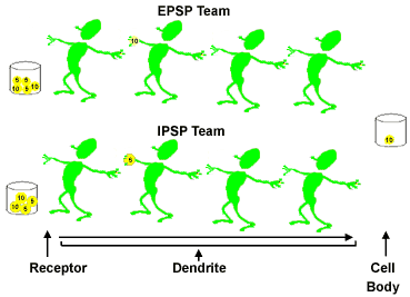
Action Potential Game
Game designed by Jessica Koch
Objective: Race to raise the resting potential in a higher place threshold to fire an action potential.
Groundwork: When neurotransmitters cross a synapse, they can bind with receptors on dendrites. This binding tin issue in a modify in the electric potential of a neuron. An excitatory postsynaptic potential occurs with the neuron becomes depolarized, raising the electrical potential from its baseline of about -70 mV and bringing information technology closer to threshold and increasing the take chances that an activeness potential will burn. An inhibitory postsynaptic potential occurs when the electrical potential is lowered, making it less likely an action potential will be generated. If the electrical potential is raised so that information technology reaches the threshold, an activeness potential will fire down the axon of a neuron.
How to Play: Players should be divided into two teams: the Excitatory Postsynaptic Potential (EPSP) Squad and the Inhibitory Postsynaptic Potential (IPSP) Squad. The teams volition race to see who can go the greatest point to their squad's prison cell body in 30 seconds. Each squad lines upwards to human activity like a dendrite. A betoken, (a modest ball), is passed from person to person much like how an electrical signal travels down a dendrite toward the cell body. Each EPSP team betoken successfully transferred to the cell torso is worth +5 or +10 mV (millivolts); each IPSP Team betoken is worth -5 or -x mV. The signals are passed down the dendrites until they achieve the end and are tossed into the cell trunk container. Only one signal ball can exist passed at a fourth dimension significant that a dendrite must drop the ball (point) into the prison cell trunk container before the first person in the dendrite can pass the next ball (bespeak).To Win: The typical resting potential of a neuron is -70 mV. To cause an activity potential the membrane potential must reach -55 mV. Therefore at the terminate of 30 seconds the signals are summed from the cell body container. The total amount of millivolts is added to -70 mV to see if an action potential is fired. If an activity potential is fired the EPSP squad wins! If not then the IPSP team wins!
Materials:
- 3 large containers or tupperware
- Nearly 32 ping pong balls, labeled with black marker -5, +5, -10, +x (8 of each). Each brawl should also exist labeled with the squad name: EPSP or IPSP.
Nervous System Child
It's a bird, information technology's a airplane....no it's "Nervous System Kid" (also known as "Brain Boy" or "Gyri Girl")! Become a large slice of butcher paper - large plenty for a student to lie down on. Have a educatee lie down on this paper and outline his or her body. Now fill-in and color this outline with parts of the nervous organization or employ the pictures of the organs supplied below. The brain and spinal string should be easy. Don't forget the sense organs (optics, ears, oral fissure, nose, skin). Follow a diagram of the peripheral fretfulness to add more features to your drawing. Also, characterization the structures that are drawn.
Materials:
- Butcher paper
- Markers (to outline and color the picture)
- Pens and pencils (to label the structures)
- Pictures of internal and sense organs - cut out, paste on your body outline and color (use the "back" push of your browser to bring you dorsum to this page):
- Brain
- Olfactory organ
- Eyes
- Oral fissure
- Ears
- Heart/Lung
- Digestive Arrangement
Mr. Egghead - The Cerebrospinal Fluid
 The cerebrospinal fluid (CSF) has several functions. One of these functions is to protect the encephalon from sudden impacts. To demonstrate how this works, we need to bring in "Mr. Egghead." Mr. Egghead is a raw egg with drawn-on face. The inside of the egg represents the brain and the egg shell represents the pia mater (the inner most layer of the meninges or coverings of the brain).
Put Mr. Egghead in a container (tupperwear works fine) that is a bit larger than the egg. The container represents the skull. Now put a tight peak on the container and shake it. Yous should observe that shaking the "encephalon" (the egg) in this state of affairs results in "harm" (a broken egg).
Now repeat this experiment with a new Mr. Egghead, except this time, fill the container with h2o. The water represents the cerebrospinal fluid. Note that shaking the container does not cause the "brain damage" equally earlier because the fluid has cushioned the brain from injury.
You could brand this into a scientific discipline fair projection: exam the hypothesis that "The cerebrospinal fluid and skull protect the encephalon from touch injury." Drib Mr. Egghead from a standard height (or heights) in different conditions: one) with fluid in the container, 2) without fluid in the container, 3) with different fluids or materials (sand, rocks) or 4) in unlike shaped containers, etc. Brand sure yous keep notes to record your observations!
Materials:
- Eggs (at least 2)
- Markers to draw on a face (waterproof)
- Plastic container with summit.
- H2o (to fill the container)
Piece and Dice - Learning Directions and Planes of Section
I way to learn the planes of sections and anatomical directions is to model the brain with fruit. That's right, fruit....the bigger the better...a melon (honey dew or cantaloupe) works nicely. Make eyes, a nose, ears and a mouth out of cork and stick them on the melon head with toothpicks. Or better withal, get a set of "Mr. Potato Head" body parts and stick them into the melon. The optics, olfactory organ, ear and rima oris requite a sense of "which way is the front" to the round melon. Now make your sections with a large knife...a coronal (frontal) department commencement, then a horizontal department, so a sagittal section. Run across the "slice page" for the correct directions and planes.
Materials:
- A melon - a honey dew or cantaloupe work
- Cork or Mr. Potato Head trunk pieces
- Knife - to cutting melon


Emotion Notion
How many emotions do y'all have? Happy, sad, mad, surprised? Brand an "Emotion Collage" past cutting out mag pictures of people expressing different emotions. Mucilage the pictures on a piece of newspaper or brand a poster to show the dissimilar emotions. You could make separate papers or posters of different emotions.
Materials:
- Magazines with pictures of people
- Scissors
- Glue
- Paper or poster board
Brain Comparisons
How is your brain similar to other objects? For example, how is your brain like a bowl of Jell-O? How is information technology different? Are they both soft? Practice they take layers? Can they shop information? Practise they employ electricity? Do they comprise chemicals? Give each person a different object. Each person must brand a list of similarities and differences betwixt their object and a brain.
Materials:
- Suggested objects: Jell-O, tape recorder, balloon, apple tree, camera, computer, telephone, volume, brawl.
Brain Charades
Although information technology's non as well hard to describe what the brain does, it's not too easy to human action it out. Effort to draw the functions of the brain and nervous system with this game of "Brain Charades."
Write down words that depict brain functions on small pieces of newspaper. This table of words will help you lot go started:
| Vision | Smell | Taste | Touch | Hearing |
| Emotions | Motion | Memory | Speech | Heart Rate |
| Breathing | Thinking | Planning | Problem Solving | Reading |
| Control Hormones | Slumber | Remainder | Eating | Drinking |
Mix the papers in a bowl, bag or a hat. A role player should selection a paper out of the basin then human activity out the function. Anybody else should attempt to guess what the player is interim out. Actors must remain silent. When someone guesses the action, write the discussion on the lath. Another thespian should select a new word and deed it out. Repeat the game until all of the words have been identified correctly.
Materials:
- Paper
- Pen or pencil
- Container for words
| Get TO: | Hearing | Odor | Taste | Bear on | Vision | Working Together |

How To Make A Neuron Model,
Source: https://faculty.washington.edu/chudler/chmodel.html
Posted by: farrquir1968.blogspot.com
0 Response to "How To Make A Neuron Model"
Post a Comment